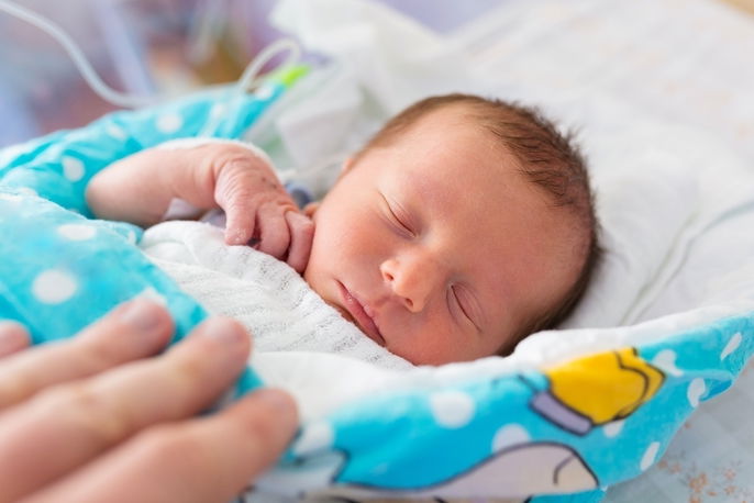
Retinopathy of prematurity (ROP) is a condition affecting premature newborns and involves abnormal growth of blood vessels in the retina, which is the part of the eye that detects light and sends signals to the brain via the optic nerve, forming images.
ROP does not usually cause any symptoms, and is most often detected on routine exam by a neonatologist in the NICU (neonatal intensive care unit). This condition affects premature babies born before 30 weeks of pregnancy or with a birthweight less than 1500 grams.
The majority of newborns with ROP will have a good outcome without serious complications, however a doctor may recommend laser treatments, injections of bevacizumab, or surgery in the event of an increased risk for retinal detachment.

Common symptoms
Retinopathy of prematurity does not cause symptoms. It is most often detected on fundoscopic exam (an eye exam that looks at the back of the eye) by an ophthalmologist in the NICU.
At more advanced stages, ROP can cause retinal detachment, which can present with the following symptoms:
- Eye twitching or other unusual eye movements;
- Difficulty tracking objects;
- White pupils.
It is important that ROP is identified as early as possible, as retinal detachment can lead to vision loss or blindness.
Confirming the diagnosis
The diagnosis of retinopathy of prematurity is made by an ophthalmologist based on a fundoscopic eye exam using eye drops to dilate the pupil and observe the back of the eye where the retina is found.
This type of exam is done for all babies with a birthweight less than or equal to 1500 grams and those born before 30 weeks gestation. The first exam for ROP is done around 30 days after birth and is repeated every 1 to 3 weeks, or as indicated by a neonatologist.
Possible causes
Retinopathy of prematurity is caused by the incomplete development of blood vessels in the retina in babies born before 30 weeks gestation or with a birthweight less than or equal to 1500 grams.
This is because the normal development of blood vessels in the retina begins around the 14th week of pregnancy and does not end until after the baby is born at term.
Normal development of these blood vessels may stop in babies who are born very early and receive supplemental oxygen in the NICU. Instead, abnormal blood vessels can develop that may grow in the wrong direction, increasing the risk for retinal detachment that can lead to blindness.
The earlier the baby is born, the higher the risk of developing ROP. External factors like lights or camera flashes do not affect risk.
Treatment options
In the majority of cases, retinopathy of prematurity does not require any specific treatment apart from maintaining regular follow up with a neonatologist.
If there are concerns for an increased risk of blindness, the neonatologist may refer the baby to an ophthalmologist, who can arrange for the most appropriate treatment. Different treatment options include:
- Laser photocoagulation: a form of treatment using laser beams to stop the growth of abnormal blood vessels that pull the retina out of place. It is most often used when ROP is diagnosed early;
- Anti-vascular endothelial growth factor (anti-VEGF) therapy: these medications are given via intravitreal injection (an injection given directly into the baby's eye) in order to interrupt the abnormal growth of blood vessels in the retina;
- Scleral buckling: this method is used in cases of advanced retinopathy when the retina is already starting to detach from the back of the eye. It involves placing a small band around the eyeball to keep the retina in place;
- Vitrectomy: a surgery used in the most advanced cases to remove vitreous gel and scar tissue from the interior of the eye and replace it with a transparent substance.
Following treatment, the baby may need to use an eye patch, especially in the case of vitrectomy or placement of a surgical band.
Recovery time
Following treatment for retinopathy of prematurity, the baby will need to remain in the hospital for at least one day, or until the effects of the anesthesia have completely worn off.
During the first week following surgery, the parents or caregivers should apply prescription eyedrops daily in order to prevent an infection from developing that could affect the results of the surgery or make the problem worse.
Babies diagnosed with ROP should be followed by an ophthalmologist until the condition is considered to be cured. Repeat evaluations may be done while the baby is still in the hospital and after discharge.
In some cases, the baby will need surgery after they are discharged from the NICU, which will require readmission at the time of surgery.


